Written and clinically reviewed by Dr. Mehmood Asghar, BDS, MPhil, PhD. Last reviewed: October 10, 2026. See our editorial standards.
Quick answer
White spots on the gums are often caused by irritation, a canker sore, surface buildup, thrush or healing tissue. Appearance alone cannot confirm the cause. Arrange a dental examination if a patch cannot be wiped away, feels thick or hard, grows, bleeds, keeps returning or remains for more than two weeks. Seek urgent care for rapidly spreading facial swelling, fever with worsening pain, uncontrolled bleeding, or difficulty breathing or swallowing.
Try not to repeatedly scrape or check the area. Instead, note whether it wipes away gently, hurts, changes size, or followed whitening, brushing, an appliance or dental treatment. Those details are more useful to a dentist than colour alone.
If the white area is immediately around an implant or healing cap, see our focused guide to white tissue around a dental implant. If it appeared after grafting, use our dental bone graft guide and contact the treating clinician if recovery is worsening.
What to do now: red, amber or green?
| Level | What you notice | What to do |
|---|---|---|
| Red — urgent | Difficulty breathing or swallowing, rapidly spreading facial swelling, uncontrolled bleeding, or feeling seriously unwell. | Seek urgent dental or medical care now. |
| Amber — book an examination | A non-wipeable, thick, hard, rough, red-and-white, painful, bleeding, growing or recurring patch; pus or a bad taste; or an unexplained area lasting more than two weeks. | Arrange a prompt dental examination. Do not scrape or self-diagnose it. |
| Green — monitor briefly | A small area after minor trauma, whitening or recent dental work that is already improving and has no warning signs. | Use gentle oral care and monitor it. Book a dentist if it stops improving or persists. |
What pattern do you notice?
A white spot, a wipeable film and generally pale gums are not the same problem. This comparison can help you describe the change clearly, but it is not a diagnosis.
| Pattern | Possible explanations | Next step |
|---|---|---|
| Soft film that wipes away gently | Plaque, food debris, dry-mouth residue or oral thrush | Use gentle oral hygiene. Book an examination if it returns, spreads or leaves sore red tissue. |
| Painful white or yellow centre with a red border | Canker sore or minor trauma | Monitor healing. Seek care if large, severe, recurrent or present beyond two weeks. |
| White area beside a tooth with swelling, bleeding, pus or bad taste | Gum inflammation or a dental infection | Arrange prompt dental care, especially if pain or swelling is increasing. |
| Thick, rough or non-wipeable patch | Frictional change, leukoplakia, oral lichen planus or another lesion | Book a professional examination rather than trying to remove it. |
| Gums look pale throughout the mouth | A general health issue may be involved | Discuss it with a dentist or doctor, particularly with fatigue, dizziness or breathlessness. |
Common causes of white spots on gums
1. A wipeable white film: plaque, residue or thrush
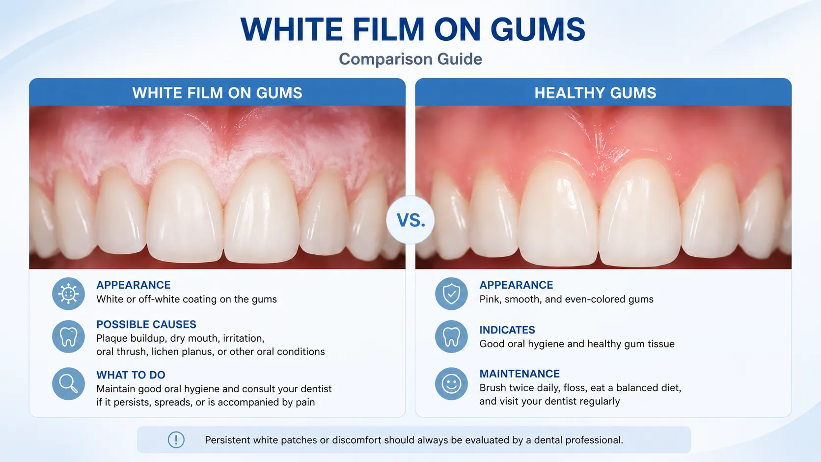
A white film on the gums may be surface buildup, food debris, dry-mouth residue or shedding of irritated surface cells. Plaque often collects along the gumline, especially around crowded teeth, braces, retainers, crowns or areas that are difficult to clean. Gum disease itself more often makes gums red, swollen or prone to bleeding than white.
Oral thrush can cause creamy white patches on the gums and elsewhere in the mouth. They may wipe away and leave red or sore tissue. The CDC lists white patches, soreness, a cotton-like feeling, altered taste and pain while eating or swallowing among possible symptoms. Thrush is more likely after antibiotics, with inhaled corticosteroids, dentures, diabetes, dry mouth or a weakened immune system. A dentist or doctor can confirm it and prescribe antifungal treatment when appropriate.
2. A painful white or yellow sore: canker sore or trauma
Canker sores often look round or oval with a white or yellow centre and a red border. They are not contagious. Minor sores commonly heal within one to two weeks, but unusually large, very painful, recurrent or slow-healing sores should be checked. Possible triggers include biting the gum, sharp food, dental work, braces, aggressive brushing and stress. A rough tooth, filling, denture or appliance can also repeatedly rub the tissue.
A white or peeling area can also follow a burn from hot food, whitening gel, strong mouthwash or another chemical irritant. Stop the suspected irritant and use gentle care. Never place aspirin, bleach, undiluted peroxide or essential oils directly on the gums.
3. A white area around a tooth: buildup, irritation or infection
People often ask, “Why are my gums white around my teeth?” A thin line that brushes away gently may be plaque or debris. Temporary whitening after bleaching treatment can be chemical irritation or tissue blanching. A sore beside a sharp tooth or appliance may be traumatic.
Gum disease is not usually diagnosed by a white spot. Look instead for bleeding, swelling, tenderness, recession, bad breath, loose teeth or discomfort when chewing. The CDC explains that periodontal disease involves inflammation and infection of the tissues supporting the teeth. A pimple-like white bump with swelling, throbbing pain, pus or a bad taste may be a drainage point from a dental infection and needs prompt care.
4. A thick patch that does not wipe away
A persistent thick, rough or non-wipeable white patch needs professional assessment. Possible causes include friction from a rough tooth or denture, leukoplakia, oral lichen planus and other oral lesions. MedlinePlus describes leukoplakia as a white patch that cannot be scraped off. Tobacco is a major risk factor, while chronic irritation may also contribute.
Most white patches are not cancer, and seeing a dentist does not mean the worst is expected. However, some persistent changes need examination or a biopsy to identify them accurately. White-and-red areas, a lump, numbness, unexplained bleeding, neck swelling or difficulty chewing or swallowing make timely assessment especially important.
5. Lacy white lines or sore red-and-white areas
Oral lichen planus can appear as lacy white lines, red swollen tissue or open sores on the gums and other parts of the mouth. Some people have no discomfort; others notice burning, sensitivity to spicy or acidic foods, or bleeding during brushing. It is not contagious, but a dentist or oral medicine clinician should diagnose and monitor it.
6. Pale gums rather than a local spot
Gums that look pale throughout the mouth are different from a local white patch. Anaemia or another general health issue may be involved. If the change appears with fatigue, dizziness, weakness, fainting or shortness of breath, contact a healthcare professional. Sudden severe pallor with serious illness needs urgent medical care.
When should you see a dentist?
Book a dental examination if the area:
- lasts more than two weeks or keeps returning;
- cannot be wiped away gently;
- feels thick, hard, raised or rough;
- is growing, bleeding or mixed with red tissue;
- comes with pain, swelling, pus, a bad taste or fever;
- appears with gum recession, a loose tooth or difficulty chewing;
- is associated with numbness, a neck lump, unexplained ear pain or difficulty swallowing; or
- is present in someone who uses tobacco or drinks heavily.
The National Institute of Dental and Craniofacial Research advises having an unexplained white or red patch, sore, lump, thick area, bleeding, numbness or swallowing difficulty assessed if it lasts longer than two weeks.
Seek urgent dental or medical care for rapidly spreading facial swelling, trouble breathing or swallowing, uncontrolled bleeding, fever with worsening pain, or signs that an infection is spreading.
How a dentist works out the cause
Your dentist will ask when the change began, whether it hurts or wipes away, whether it has changed, and about recent dental treatment, appliances, medicines, dry mouth, diabetes, antibiotics, smoking and alcohol. They will examine the gums, teeth, tongue, cheeks, palate, floor of the mouth, restorations and neck.
Further tests depend on the findings. A gum assessment and X-rays may be useful if periodontal disease or a dental infection is suspected. A swab or small scraping may help identify thrush. A persistent or unusual non-wipeable patch may be referred to an oral medicine clinician, oral surgeon or ENT specialist for a biopsy. A biopsy is a diagnostic step; it does not by itself mean cancer is present.
Treatment depends on the diagnosis
| Likely cause | Possible professional care |
|---|---|
| Plaque or gingivitis | Professional cleaning, improved brushing and interdental cleaning, and follow-up. |
| Periodontitis | Gum measurements, X-rays, deep cleaning and an individual maintenance plan; surgery is sometimes needed. |
| Oral thrush | Antifungal treatment and attention to contributing factors such as dentures, inhalers, diabetes or dry mouth. |
| Canker sore or trauma | Removal of the irritant, pain relief or topical treatment; recurrent or persistent ulcers may need further investigation. |
| Leukoplakia or another persistent patch | Removal of irritants, tobacco cessation support, monitoring, specialist referral or biopsy when indicated. |
| Oral lichen planus | Monitoring and symptom control, sometimes with prescription medication. |
What you can safely do while waiting
- Brush gently twice a day with fluoride toothpaste and a soft-bristled toothbrush.
- Clean between the teeth once daily without forcing floss into a sore area.
- Stay hydrated and avoid foods that sting the area.
- Pause a whitening product or harsh mouthwash if it appears to have caused irritation.
- Avoid tobacco, vaping and direct chemical “remedies.”
- Do not scrape, pick or repeatedly press the patch.
- Take one clear photo every few days in similar lighting and note whether it is changing.
For anxiety, use a simple monitoring rule: check no more than once daily, record one observation, and act on the red or amber signs above. Repeated checking can increase distress without clarifying the cause. If mouth sensations are becoming hard to ignore, our guide to dental anxiety and symptom hyperawareness explains why this happens.

Frequently asked questions
Why are my gums white around my teeth?
A wipeable line may be plaque or debris; a temporary white area after whitening may be irritation; and a painful spot may be a sore. White tissue with bleeding, swelling, pus or a bad taste needs dental assessment. A thick area that does not wipe away should also be checked.
Is white plaque on the gums a sign of gum disease?
Surface plaque can collect at the gumline and contribute to gingivitis, but gum disease usually causes redness, swelling and bleeding rather than a distinct white patch. If the material does not brush away gently, or the gums are painful or swollen, see a dentist.
Can white spots on gums be cancer?
Most are not cancer. Because a persistent white or red patch can occasionally be a warning sign, arrange an examination if it lasts more than two weeks, cannot be wiped away, grows, bleeds or feels thick or rough.
Should I scrape a white patch off my gum?
No. Gentle brushing is reasonable, but forceful scraping can injure the tissue, cause bleeding and make the area harder to assess. A true lesion will not be treated by scraping.
How long should I wait before getting it checked?
Do not wait if there is spreading swelling, fever, pus, severe pain, bleeding or difficulty breathing or swallowing. For an unexplained patch without urgent symptoms, arrange an examination if it is not clearly improving or remains after two weeks.
Bottom line
White spots on gums have many possible causes, and colour alone cannot diagnose them. A small area after minor irritation that is already improving can usually be monitored briefly. A persistent, non-wipeable, thick, rough, growing, bleeding or red-and-white patch should be examined. Seek urgent help for spreading swelling, fever with worsening pain, uncontrolled bleeding, or breathing or swallowing difficulty.
Medical disclaimer
This article is for education and does not replace an examination or personalised dental or medical advice. If you are worried about a change in your mouth, contact a qualified dentist or healthcare professional.
Sources
- National Institute of Dental and Craniofacial Research: Oral Cancer
- CDC: About Periodontal Disease
- CDC: Candidiasis Signs and Symptoms
- American Dental Association: Periodontitis
- Mayo Clinic: Canker Sore
- Mayo Clinic: Oral Thrush
- Mayo Clinic: Leukoplakia
- MedlinePlus: Leukoplakia
- Mayo Clinic: Oral Lichen Planus

Dr. Mehmood Asghar is a Dentist and an Assistant Professor of Dental Materials, with nearly a decade of clinical experience. He holds a Ph.D. in Dental Biomaterials from Universiti Malaya and a Bachelor of Dental Surgery (BDS) from NUST. As a dedicated researcher with over 25 peer-reviewed publications and more than 310 citations in international journals, Dr. Asghar bridges the gap between clinical dentistry and advanced biomaterials research to provide patients with evidence-based, practical dental advice.
Discover more from The Calm Dentist Blog
Subscribe to get the latest posts sent to your email.