Written and clinically reviewed by Dr. Mehmood Asghar, BDS, MPhil, PhD. Last reviewed: August 2, 2026. See our editorial standards.
Quick Answer: Dental Bone Graft Stitches Fell Out
If your stitches came out after a dental bone graft, it does not necessarily mean the graft came out or failed. Dissolvable sutures can loosen during healing, and a small amount of graft material, fibrin, or collagen membrane may remain attached when a stitch comes away.
Contact your dentist or oral surgeon promptly if the wound opens, a large piece of material comes out, or you develop persistent bleeding, worsening swelling, severe pain, pus, fever, or a bad taste. Do not push anything back into the socket or repeatedly touch the area.
Until then:
-
- Do not try to push the material back into the socket.
-
- Avoid touching the area with your tongue or fingers.
-
- Continue using any prescribed mouth rinse exactly as instructed.
-
- Eat soft foods and avoid chewing on the surgical site.
-
- Call your dentist or oral surgeon if you’re unsure what came out.
In most cases, losing a few particles attached to a stitch doesn’t mean your bone graft has failed
If you noticed a small white piece attached to your stitches after oral surgery, your first thought was probably, “Did my bone graft just come out?”
It’s one of the most common concerns patients have after a dental bone graft, and understandably so. Seeing unfamiliar material in your mouth after surgery can be alarming, especially when you’ve invested time and money into preparing for a future dental implant.
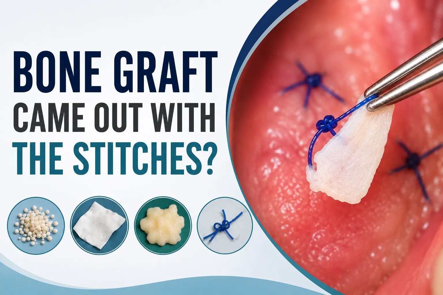
The good news is that material attached to your stitches is often not your actual bone graft. In many cases, it’s a dissolvable collagen membrane, dried blood clot, fibrin, or even the suture itself. Even if a few tiny bone graft particles come away with the stitches, it doesn’t automatically mean the graft has failed.
This guide will help you identify what you may be seeing, explain why it sometimes happens, and help you understand when it’s worth contacting your dentist.
When Should Bone Graft Stitches Come Out?
Dissolvable stitches soften and disappear during healing, but the timing varies. NHS oral surgery guidance notes that some may fall out within a few days while others take up to three weeks to dissolve. This is general oral-surgery guidance, not a deadline for every bone graft. Follow the timing your surgeon gave you.
Non-dissolvable stitches need a removal appointment. If you are unsure which type you have, or a stitch comes out earlier than your surgeon expected, contact the treating office. Do not pull or cut remaining stitches yourself.
What to Tell Your Dentist When a Stitch Comes Out
Tell the office when surgery took place, when the stitch came out, whether one or several stitches are missing, and whether the gum edges have opened. Describe any material lost and whether pain, swelling, bleeding or discharge is improving or worsening. Do not pull the cheek or probe the wound to investigate.
If your main concern is loose material, see our guide to bone graft particles and membrane loss. For worsening symptoms, see dental bone graft infection signs and contact your treating clinician.
Why Does Material Sometimes Come Out With the Stitches?
A bone graft isn’t simply placed into an empty hole and left exposed.
Instead, your oral surgeon carefully layers several components together:
-
- Bone graft material
-
- Blood clot
-
- Healing tissue
-
- A protective collagen membrane (in many cases)
-
- Sutures to hold everything in place
These layers gradually integrate with your own tissues through the bone graft healing stages.
When stitches are removed—or dissolve naturally—they may occasionally bring some of the surface material with them.
This doesn’t necessarily mean the graft underneath has been disturbed. Many oral surgeons use a collagen membrane to protect the graft while the surrounding tissues heal. This technique, known as guided bone regeneration, is widely accepted in modern implant dentistry.
Think of it like removing the protective dressing from a healing wound. Sometimes the dressing sticks slightly to the surface while the deeper tissue remains intact.
Was It Actually Bone Graft Material?
Most patients assume anything white equals bone graft.
In reality, several materials can look remarkably similar.
| What Came Out? | Appearance | Should You Worry? |
|---|---|---|
| Bone graft particles | Small white or beige grains that resemble coarse sand | Usually not if only a few particles are lost |
| Collagen membrane | Thin white sheet or sponge-like material | Contact your treating team if the membrane comes away; the timing and graft type matter |
| Fibrin | Soft white or yellow coating | Normal healing tissue |
| Blood clot | Dark red or brown jelly-like tissue | May require review if lost early after surgery |
| Dissolvable stitch | White, tan, or purple thread | May occur as it dissolves; seek advice if earlier than expected or the wound opens |
Without examining the area, even dentists often cannot identify the material from a patient’s description alone.
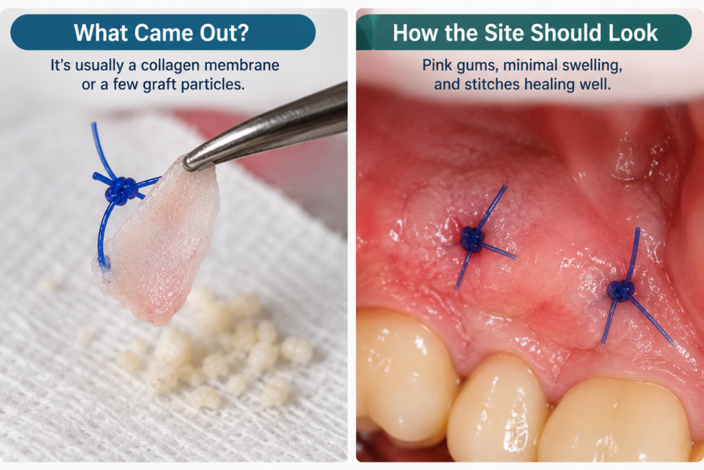
How to Tell the Difference
1. Bone Graft Particles
Bone graft materials are commonly made from:
-
- Processed human donor bone
-
- Bovine (cow) bone
-
- Synthetic calcium phosphate
-
- Bioactive ceramic materials
These materials look like tiny grains of coarse salt or sand.
You may occasionally notice one or two particles on your gauze while changing it during the first few days after surgery.
This is usually considered normal.
The graft is packed with hundreds—or sometimes thousands—of microscopic particles.
Losing a few surface granules rarely affects healing.
2. Collagen Membrane
Many dentists place a collagen membrane over the bone graft before closing the gums.
Its purpose is to:
-
- Keep the graft stable
-
- Prevent soft tissue growing into the graft
-
- Protect the healing bone
-
- Improve regeneration
Collagen membranes gradually dissolve over several weeks.
Occasionally, part of the membrane becomes exposed or separates as the stitches loosen.
Patients often mistake this white sheet-like material for the bone graft itself.
Unlike graft particles, membranes usually appear:
-
- Flat
-
- Soft
-
- Flexible
-
- Sponge-like
Rather than gritty.
3. Fibrin (Normal Healing Tissue)
A white or yellow film covering the surgical site is often fibrin, which is a normal part of wound healing—not an infection. The American Dental Association (MouthHealthy) explains that healing tissues can look unusual during the first few weeks after oral surgery.
Fibrin forms naturally after surgery and appears as a white, yellow, or cream-colored coating over the wound.
Many patients worry they have:
-
- pus
-
- infection
-
- exposed bone
-
- failed graft
when they’re actually seeing completely normal healing tissue.
Unlike infection, fibrin usually:
-
- doesn’t smell
-
- isn’t painful
-
- doesn’t produce swelling
-
- gradually disappears on its own
Removing it or trying to clean it away can delay healing.
4. Blood Clot
Immediately after surgery, a blood clot forms over the graft.
This clot protects the underlying bone while healing begins.
If the clot is dislodged within the first few days, healing may become slower and discomfort may increase.
Fortunately, blood clots look very different from bone graft material.
They’re typically:
-
- dark red
-
- purple
-
- jelly-like
-
- soft
rather than white and gritty.
Why Bone Graft Particles Sometimes Stick to Stitches
As your tissues heal, tiny graft particles near the surface can become incorporated into the healing tissue surrounding the sutures.
When the stitches loosen or are removed, those superficial particles may come away together.
This doesn’t mean the entire graft has detached.
Remember that successful bone graft healing depends on the bulk of the graft remaining stable beneath the gum tissue, not on every individual particle staying perfectly in place.
Many surgeons even tell patients to expect seeing a few particles during the first week after surgery.
What If the Stitches Came Out but the Bone Graft Stayed in Place?
If the stitches came out but the gum edges remain mostly together and only a few small particles were lost, the deeper graft may still be protected. Sutures support the wound during early healing, but they are not the graft itself. A loose or missing stitch does not prove that the entire graft has been displaced.
Contact your dentist or oral surgeon if the stitches come out earlier than expected, particularly during the first few days. They can check whether the wound is still closed, whether a membrane is exposed, and whether monitoring or treatment is needed. Do not cut remaining stitches or try to push loose material back into the area.
Does This Mean My Bone Graft Failed?
Usually, no.
A successful bone graft isn’t judged by whether a few particles are lost.
Instead, dentists look for signs such as:
-
- healthy gum healing
-
- absence of infection
-
- stable underlying graft
-
- new bone formation on future X-rays or CBCT scans
Losing a small amount of superficial material is rarely enough to cause failure.
However, larger losses can be more significant.
You should contact your dentist if:
-
- a large amount of graft material suddenly falls out
-
- the surgical site opens completely
-
- you develop severe pain
-
- swelling increases after initially improving
-
- pus or a bad taste develops
-
- you develop fever
If you’re concerned that the graft itself may have come out, read our complete guide on Dental Bone Graft Falling Out, which explains the warning signs, treatment options, and when emergency care may be needed.
What Will Your Dentist Look For?
If you’re unsure whether the material attached to your stitches was actually part of the bone graft, the best next step is to let your dentist or oral surgeon examine the area. In many cases, they can determine whether the site is healing normally with a simple visual examination. Occasionally, they may take an X-ray or CBCT scan if they’re concerned that a significant amount of graft material has been lost.
Rather than focusing on what came out, your dentist is usually more interested in how the surgical site looks today.
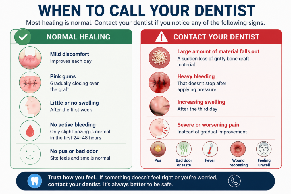
A healthy healing bone graft typically has:
-
- Pink gums that are gradually closing over the graft
-
- Mild tenderness that improves each day
-
- Little or no swelling after the first week
-
- No active bleeding
-
- No pus or unpleasant odor
These signs are much more important than finding a few white particles on your gauze or attached to a dissolving stitch.
What Happens If Some Bone Graft Material Was Lost?
Even if a small amount of graft material came away with the sutures, it doesn’t necessarily affect the overall outcome.
Bone grafts are intentionally slightly overfilled during surgery because dentists know that a small amount of remodeling and particle loss is normal. Your body gradually replaces the graft material with your own bone over several months, and not every individual particle survives this process.
Studies have shown that successful graft healing depends on maintaining enough graft volume for new bone to develop—not on preserving every microscopic granule.
In other words, losing a few surface particles is very different from losing the entire graft.
If you’re worried that a significant amount of graft material has come out, read our guide to Dental Bone Graft Falling Out, where we explain what true graft failure looks like and what treatment may be needed.
When Should You Contact Your Dentist?
Although most cases aren’t emergencies, there are situations where you shouldn’t wait until your next scheduled review.
Call your dentist promptly if you notice:
-
- A large amount of gritty graft material suddenly falls out
-
- Heavy bleeding that doesn’t stop after applying pressure
-
- Increasing swelling after the third day
-
- Severe or worsening pain or bone graft infection instead of gradual improvement
-
- Pus draining from the surgical site
-
- A persistent bad taste or foul odor
-
- Fever or feeling generally unwell
-
- The entire wound appears to have reopened
These symptoms don’t always mean the graft has failed, but they do warrant professional assessment.
Frequently Asked Questions
Is it normal for stitches to pull out bone graft particles?
Yes. Tiny graft particles can occasionally adhere to healing tissue around the sutures and come away when the stitches dissolve or are removed. Losing a few particles is generally considered normal and usually doesn’t affect the success of the graft.
What if the material was white and looked like a sponge?
This is often a collagen membrane rather than the bone graft itself.
Collagen membranes are commonly placed over grafts to stabilize the area and guide healing. As they dissolve, small pieces may become visible or detach naturally.
Can I tell whether my bone graft failed by looking at it?
Not reliably.
While patients often try to judge healing by appearance, it’s difficult to distinguish between normal healing tissue, collagen membranes, exposed graft particles, and infection without clinical training.
Your dentist may need to examine the site—and sometimes take imaging—to confirm whether healing is progressing normally.
Should I save the material that came out?
If you’re seeing your dentist within the next day or two, it can be helpful to place the material in a clean container or take a clear photograph before discarding it.
While your dentist may not need the material itself, a photo can sometimes help determine whether it appears to be graft particles, collagen membrane, or simply healing tissue.
Will my dental implant still be possible?
In most cases, yes.
Minor particle loss rarely changes the treatment plan. Once healing is complete, your dentist will assess whether enough new bone has formed before placing the implant.
Only significant graft loss or infection is likely to require additional treatment before implant placement.
The Bottom Line
Finding white material attached to your stitches after a dental bone graft procedure can be unsettling, but it doesn’t automatically mean the procedure has failed.
In many cases, the material is a dissolving collagen membrane, fibrin, or a few superficial graft particles rather than the entire bone graft. Even when small amounts of graft material are lost, the remaining graft often continues to heal successfully beneath the gum tissue.
The most important thing is to monitor how the area feels—not just how it looks. Gradually improving discomfort, healthy pink gums, and the absence of swelling or infection are all reassuring signs.
If you’re ever unsure, don’t hesitate to contact your dentist. A quick examination can usually confirm whether everything is healing as expected and provide peace of mind.
Continue Learning About Bone Grafts
If you’re recovering from a bone graft, these guides may also help:
-
- Dental Bone Graft Falling Out – Learn the difference between normal particle loss and true graft failure.
-
- Bone Graft Healing Stages – See what to expect during each phase of recovery.
-
- Signs of Bone Graft Infection – Discover which symptoms require prompt dental attention.
References
-
- American Association of Oral and Maxillofacial Surgeons (AAOMS): Dental Bone Grafting – https://myoms.org
-
- American Dental Association (ADA): MouthHealthy – https://www.mouthhealthy.org
-
- National Institute of Dental and Craniofacial Research (NIDCR): Oral Surgery and Bone Healing – https://www.nidcr.nih.gov
-
- Wang HL, Boyapati L. “PASS Principles for Predictable Bone Regeneration.” Implant Dentistry. 2006.
Disclaimer
The Calm Dentist exists to be the resource you land on instead of one that sends you spiraling. Every article here is written by dental clinicians and researchers with one goal: calm, accurate answers you can actually use. If a new symptom appears during your recovery, the post-surgical guides here are a good place to start; trusted patient-facing summaries such as WebMD’s overview of gum tissue graft surgery can also be helpful, then follow up with your surgeon for anything that concerns you. Knowing the difference between what’s normal and what isn’t is the most practical step you can take toward a successful recovery.
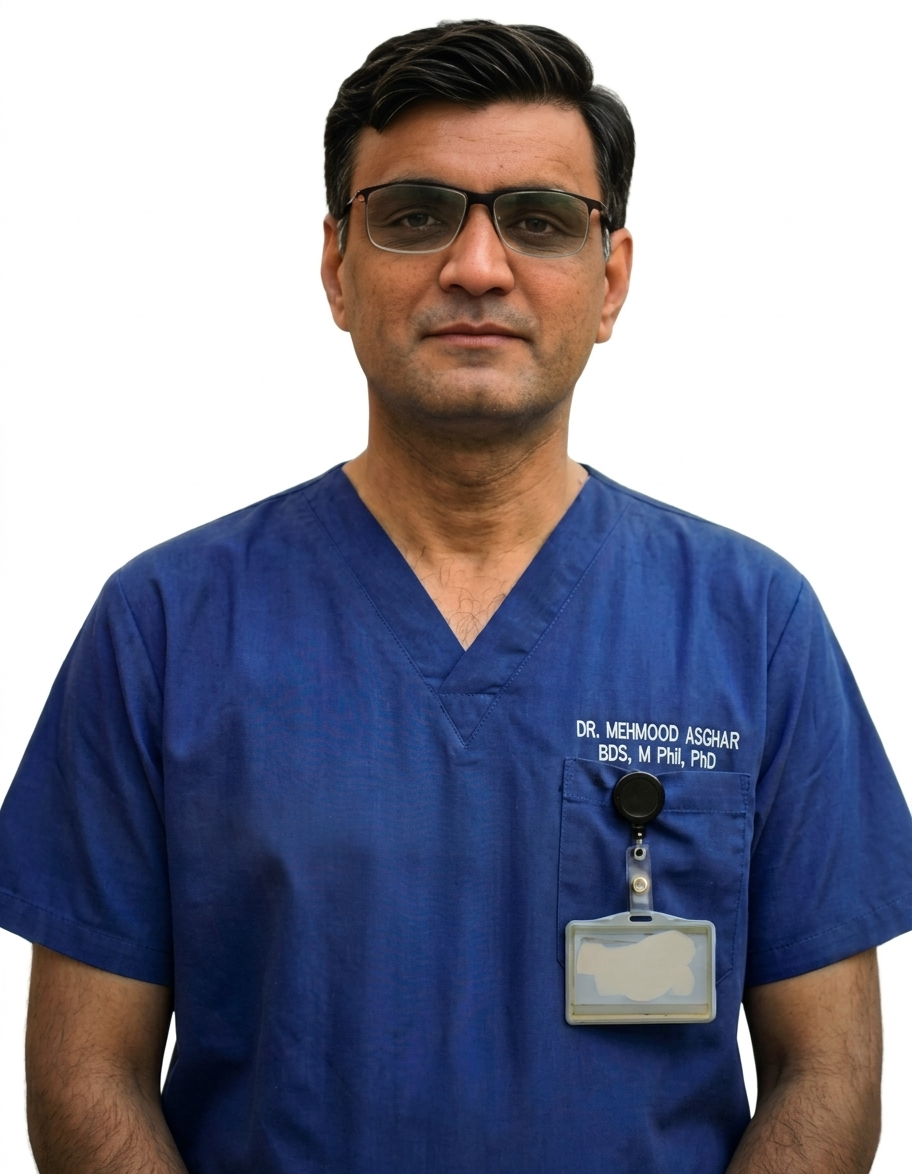
Dr. Mehmood Asghar is a Dentist and an Assistant Professor of Dental Materials, with nearly a decade of clinical experience. He holds a Ph.D. in Dental Biomaterials from Universiti Malaya and a Bachelor of Dental Surgery (BDS) from NUST. As a dedicated researcher with over 25 peer-reviewed publications and more than 310 citations in international journals, Dr. Asghar bridges the gap between clinical dentistry and advanced biomaterials research to provide patients with evidence-based, practical dental advice.
Discover more from The Calm Dentist Blog
Subscribe to get the latest posts sent to your email.